

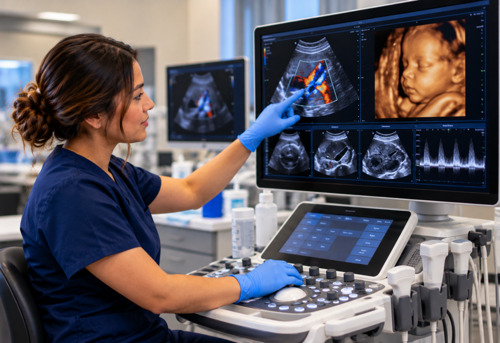
If you’re considering diagnostic medical sonography education, understanding how ultrasound technology continues to evolve can help you explore the field.
Advances in ultrasound technology affect how sound waves are transmitted, received, processed, and displayed. Depending on the application, these developments can improve image quality, support measurements, and provide additional information for clinical assessment.
Here are six established and evolving ultrasound technologies used in modern diagnostic imaging.
Ultrasound Technologies Used in Diagnostic Medical Sonography
1. Fusion Imaging Technology
Fusion imaging combines real-time ultrasound with previously acquired imaging data, such as CT or MRI scans, to help clinicians correlate anatomy across imaging methods. This technology is used for:
- Targeted biopsy guidance.
- Interventional procedures.
- Correlation of anatomical structures across imaging modalities.
Fusion imaging can support image guidance during selected diagnostic and interventional procedures.
2. 3D and 4D Ultrasound Imaging
3D ultrasound acquires volume data that can be reconstructed into three-dimensional views. 4D ultrasound displays these volumetric images over time, allowing clinicians to observe movement. This technology allows for:
- Visualization of anatomical structures from multiple perspectives.
- Additional perspectives for evaluating structural details.
- Real-time observation of movement with 4D imaging.
Advances in processing and rendering have expanded the clinical applications of 3D and 4D ultrasound, including selected obstetric and cardiac examinations.
3. Harmonic Imaging Technology
Tissue harmonic imaging uses higher-frequency echoes generated as ultrasound waves travel through tissue, rather than relying only on echoes at the original transmitted frequency.
Under appropriate imaging conditions, this technique can improve image clarity and reduce certain artifacts. Harmonic imaging supports:
- Improved definition of some tissue boundaries.
- Reduced image clutter and certain near-field artifacts.
- Improved visualization in abdominal and cardiac exams.
Tissue harmonic imaging is an established feature of many modern ultrasound systems.
4. Beamforming and Signal Processing Enhancements
Modern ultrasound systems use beamforming to coordinate transmitted pulses and combine received echoes, helping form and focus the resulting image. Enhanced beamforming allows for:
- Improved focusing across selected imaging depths.
- More consistent image quality across portions of the field of view.
- Potential improvements in contrast resolution.
These developments can improve image formation and visualization, depending on the equipment, examination, and imaging conditions.
5. Elastography Technology
Elastography estimates tissue stiffness by evaluating tissue deformation or the propagation of mechanically generated waves. It provides information about tissue mechanical properties alongside conventional grayscale imaging.
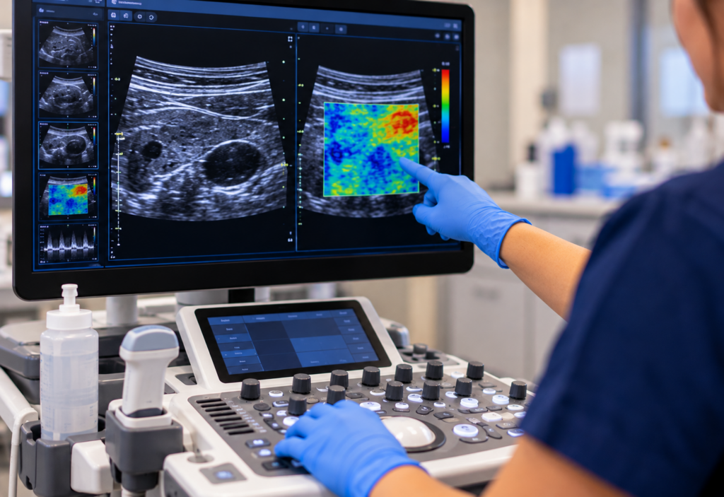
Elastography is commonly used for:
- Liver fibrosis assessment.
- Evaluating thyroid and breast lesions.
- Comparing the mechanical properties of different tissue regions.
Elastography can provide additional noninvasive information, although findings must be interpreted alongside other clinical and imaging results.
6. Automated Image Optimization and Tissue Characterization
Some modern ultrasound systems offer automated tools that adjust settings such as gain, contrast, and image processing based on the examination and imaging conditions. These technologies help:
- Support real-time image optimization.
- Help reduce certain differences in image settings between examinations.
- Support more consistent image presentation.
These tools may improve workflow efficiency, but appropriate scanning technique and professional judgment remain essential.
Building a Foundation in Sonography at Rockford Career College
Rockford Career College’s Diagnostic Medical Sonography program focuses on developing foundational knowledge and practical skills relevant to diagnostic ultrasound practice.
Students may benefit from:
- Clinical practicum experiences, as outlined in the program curriculum.
- Coursework covering anatomy, patient care, and ultrasound physics.
- Opportunities to learn ultrasound scanning techniques using program-provided equipment.
- Education relevant to sonography credentialing, with ARDMS examination eligibility determined by the applicable prerequisite pathway and requirements.
- Instruction from program faculty and access to RCC’s career services.
This combination of coursework and practical experience can help students develop foundational skills as they explore the qualifications and credentialing requirements for diagnostic medical sonography positions.
Interested in learning more? Explore Rockford Career College’s Diagnostic Medical Sonography program to review its curriculum, admissions requirements, and clinical training components.
